Integrating Technology for Precise Diagnosis
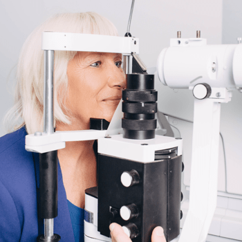
At Foresee Eyecare, it’s vital to us that we accurately diagnose your ocular conditions so we can offer personalized treatment and management strategies for your particular ocular and visual needs. We integrate technology and in-depth diagnostic equipment to detect eye diseases early and provide appropriate treatment to protect against vision loss. This allows to us to focus on preventative eye care and early detection as many eye diseases may be asymptomatic in early stages.
Book your eye exam today so we can help you take care of your eyes.
How We Use Our Technology
Your eyes are a delicate part of your body, and the inner workings of your visual system go much deeper than the naked eye can see. Our diagnostic equipment enables us to capture digital images of your eye to detect and monitor signs of eye diseases.
A Closer Look: How We Evaluate & Treat Your Eye Health
At Foresee Eyecare, we pride ourselves on our integration of technology. We invest in maintaining and updating equipment to help meet our patients’ needs.
Learn more about our technology and testing methods by exploring some of our available technologies.
RetinaLogik
RetinaLogik brings VR-powered eye exams to Foresee Eyecare. During your eye exam, we capture high-resolution images of your retina. RetinaLogik’s artificial intelligence then scans these images, detecting subtle patterns and abnormalities that may indicate conditions like diabetic retinopathy, age-related macular degeneration (AMD), and glaucoma—often before noticeable symptoms appear.
By comparing your retinal images to a vast database of clinical cases, RetinaLogik helps us identify risks early and track changes over time, allowing for personalized treatment plans and proactive care.
Eidon Ultra-Wide Retinal Photography
Using the new Eidon Ultra-Widefield retinal camera from iCare, we can observe more of your retina in a single photograph, helping us identify certain eye conditions and diseases in and around your retina.
This new technology helps us understand more about your eye health and vision than ever before, helping us address your concerns before they affect your sight.
Optical Coherence Tomography (OCT)
To get an in-depth look at your retina and its function, we use optical coherence tomography (OCT). It’s a noninvasive technology that allows us to grab high-resolution images of the retina, including its layers and thickness.
OCT technology is similar to an ultrasound, using light instead of sound to produce a clearer, sharper image of your retina.
Anterior Segment Imaging
Anterior segment imaging (ASI) enables us to take photographs and videos of the ocular structures at the front of your eye, including the orbit, eyebrows, eyelids, conjunctiva, cornea, and iris. This lets us identify and monitor conditions that may affect those parts of the eyes.
Anterior segment imaging is also valuable for monitoring and tracking healing from injuries or trauma to the front of the eye. We may share the results of this imaging with you to help explain any conditions you’re experiencing, including common ones such as dry eye and ocular surface disease.
Corneal Topography
Corneal topography is a computer-assisted diagnostic tool that allows us to determine the shape of your cornea. This helps us detect corneal conditions as we can gather a 3D map of its surface.
This technology also allows us to correctly fit specialty contact lenses and assess the eyes for laser eye surgery.
iCare Tonometer
The iCare Tonometer is a noninvasive tonometer that measures intraocular pressure. This tool is especially helpful for glaucoma screenings as it is easy, quick, and suitable for all patients.
Humphrey Visual Field Test
The Humphrey visual field test can help us detect and monitor glaucoma. It uses a centre fixation light and blinking test lights to map out your area of vision. The test can determine if certain areas in your vision are blurred or missing by appearing as grey or black areas on the results.

Refractive Errors, Myopia Control, & Vision Therapy
We use a variety of technologies to help correct vision problems and preserve your vision for years to come.
Automated Refraction Testing
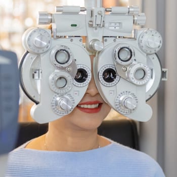
An autorefractor, or automated refractor, is a computer-controlled machine that your optometrist will use during an eye exam.
This machine observes how light changes as it enters the eye, which helps measure refractive errors, such as nearsightedness (myopia), farsightedness (hyperopia), and presbyopia. Measuring refraction can help your optometrist determine your prescription.
Medmont Meridia Topographer
Corneal topography is widely used by optometrists to describe and determine the shape of the cornea, helping them diagnose and monitor various eye conditions and diseases.
The Medmont Meridia Topographer is especially useful for myopia management and can be used to fit patients for orthokeratology and other specialty contact lenses.
Biometry & Axial Length Measurement
The Oculus Myopia Master is an all-in-one device that can noninvasively measure refraction, axial length, and corneal curvature at the same time.
These measurements help us track eye growth and changes in vision, which is a critical component to tracking myopia control and progression.
Orthokeratology (Ortho-K)
Orthokeratology, or ortho-k, is specialty contact lenses for managing myopia (nearsightedness) and other refractive errors. The custom-designed lenses are worn overnight to help improve vision by reshaping the cornea.
Farnsworth D-15 Colour Vision Test
The Farnsworth D-15 colour vision test is designed to measure the degree of colour blindness and a patient’s ability to perceive colours. This test evaluates both your type and degree of colour vision deficiency.
This test is often required to screen for candidacy for professions like firefighting and policing and offers finer details than the standard Ishihara colour vision test.
Vivid Vision: Virtual Reality
Vivid Vision is a virtual reality headset used for vision therapy. We can adding an element of fun to help train 3D vision, treating eye tracking and teaming issues like amblyopia and train 3D vision.
This noninvasive therapy has helped many children and adults succeed in their vision development.
VTS4-HoloLens
The VTS4-HoloLens comprises various exercises tailored to coordinate the eyes, promoting improved teamwork between the eyes.
The incorporation of HoloLens Augmented Reality technology introduces a diverse range of over 100 targets with varying size, detail, and retinal disparity, presented in true 3D colour. These targets, including holograms, are specifically designed to manage various vergence exercises, enhancing patient engagement.
Through the integration of these technologies and targeted exercises, the VTS4-HoloLens represents a significant advancement in vision therapy.
Dry Eye Assessments
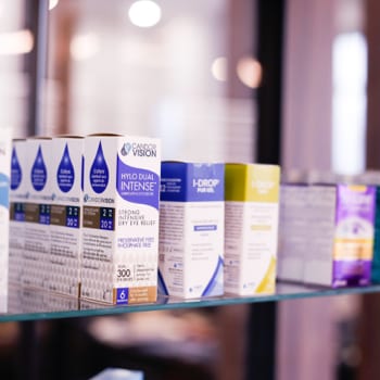
We use a mix of technologies to evaluate your eye health and tear quality, helping us tailor a dry eye treatment that works just for you. You can visit our dry eye treatment page to learn more about how we use technology to support at-home and in-office dry eye therapies.
Meibography
Meibography is the imaging of the meibomian glands (oil glands) along the edges of your eyelids.
Meibomian gland dysfunction is a condition that affects the production of your tears, which can lead to the development of dry eye. To diagnose a patient with dry eye, we may use meibography to diagnose dry eye.
Tear Film Analysis (Schirmer’s Test)
Schirmer’s test measures your tear quantity, which helps your optometrist assess the cause of your dry eye and build a recommendation to begin your treatment.
Your optometrist will place a specialty piece of paper inside of your eyelid and assess how far down the paper your tears have travelled once the time is up.
Our Brands
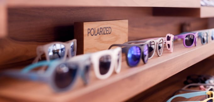
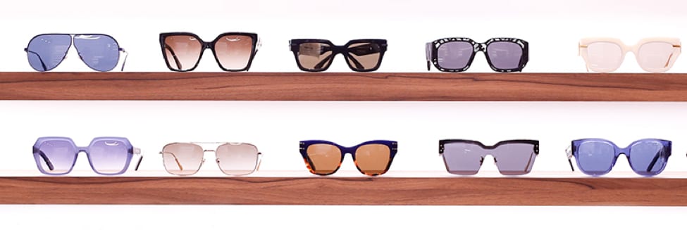
- Anne & Valentin
- Blackfin
- Carrera
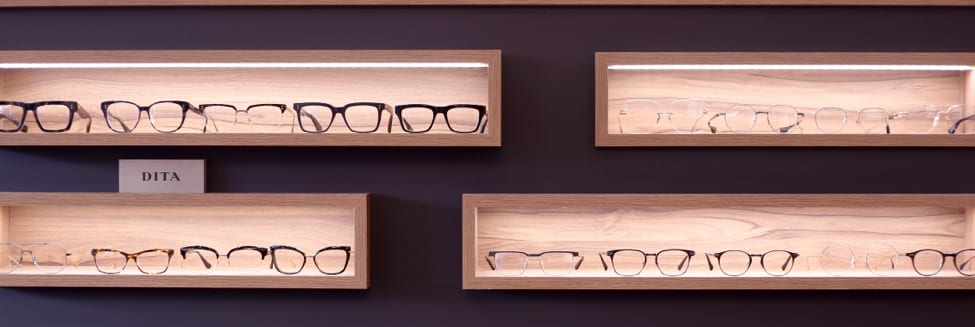
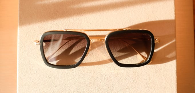
- DITA
- Dior
- Fendi
- Etnia Barcelona
- Kliik
- Lindberg
- ic Berlin
- MODO
- Maui Jim

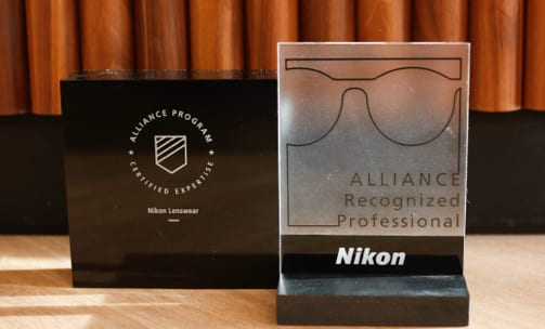

- Nano
- OTP
- OGA
- Prada
- Saint Laurent
- Ray-Ban
- Starck
- Silhouette
- Tom Ford
- Tiffany
- Tomato Glasses
- WEWE
Visit Our Location
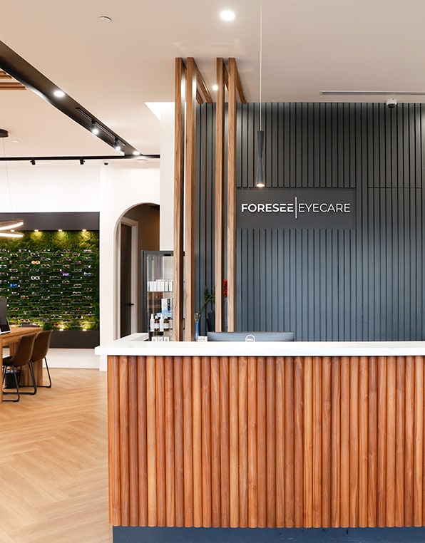
Our office can be found off of Major MacKenzie Drive West in the same plaza as the Medical Centre and Tim Hortons. The location of our office is 965 Major Mackenzie Dr. West, Units 3 & 4.
Hours of Operation
Our Address
- 965 Major Mackenzie Dr. West, Units 3 & 4
- Vaughan, ON L6A 4P8
Contact Information
- Phone: 289-553-EYES (3937)
- Fax: 289-553-3939
*Friday by appointment only.
**We are closed on the Saturdays of long weekends and holidays.
